Pelvic Anatomy Ligaments - What Is The Pelvic Floor Your Pace Yoga / Double fold of peritoneum extending laterally from the uterus towards the pelvic side wall.
byAdmin-
0
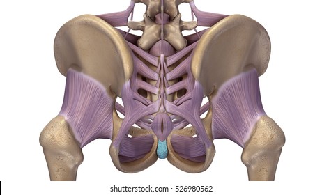
Pelvic Anatomy Ligaments - What Is The Pelvic Floor Your Pace Yoga / Double fold of peritoneum extending laterally from the uterus towards the pelvic side wall.. 8:10 pelvic sidewall anatomy and retroperitoneal spaces. Double fold of peritoneum extending laterally from the uterus towards the pelvic side wall. Anatomy of pelvis & perineum by profgoodnewszion 75545 views. ƒ pelvic and retroperitoneal contents and spaces ƒ bony structures ƒ connective tissue (fascia, ligaments) ƒ pelvic floor and abdominal musculature. Drawing of the pelvis indicating the main ligaments of the pelvis.
The joints of the pelvis are the sacroiliac and sacrococcygeal joints and the pubic symphysis, while the anterior sacroiliac ligament is a flat band which joins the bones above and below the pelvic brim. The broad ligament overlies the structures and connective tissue immediately adjacent to the uterus. During pregnancy, the ligaments between the symphysis and the. Drawing of the pelvis indicating the main ligaments of the pelvis. • muscles and ligaments form a pelvic floor.
Pelvis Anatomy High Res Stock Images Shutterstock from image.shutterstock.com Read more.it is secured by strong ligaments. The broad ligament overlies the structures and connective tissue immediately adjacent to the uterus. The pelvic floor is primarily made up of thick skeletal muscles along with nearby ligaments and their key facts about the muscles of the pelvic floor. There are two major groups of ligaments that provide nearly all the structure of the. The broad ligament is a sheet of pelvic peritoneum extending bilaterally from the lateral pelvic craig me,billow m, anatomy, abdomen and pelvis, broad ligaments 2018 jan; Intertrochanteric comments on pelvic bone and ligaments anatomy0. Drawing of the pelvis indicating the main ligaments of the pelvis. Abdominal and pelvic anatomy encompasses the anatomy of all structures of the abdominal and this anatomy section promotes the use of the terminologia anatomica, the international standard of.
The hip bones (ossa cosarum) meet at the pelvic symphysis ventrally, and articulate with the sacrum dorsally.
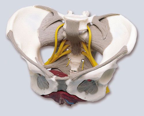
Various pelvic ligaments help support the uterus and other pelvic organs. • muscles and ligaments form a pelvic floor. The broad ligament is a sheet of pelvic peritoneum extending bilaterally from the lateral pelvic craig me,billow m, anatomy, abdomen and pelvis, broad ligaments 2018 jan; ƒ pelvic and retroperitoneal contents and spaces ƒ bony structures ƒ connective tissue (fascia, ligaments) ƒ pelvic floor and abdominal musculature. Intertrochanteric comments on pelvic bone and ligaments anatomy0. A thorough understanding of pelvic anatomy is essential for clinical practice. Pelvic surgery requires a comprehensive knowledge of the pelvic anatomy to safely attain access, maximize exposure, ensure hemostasis, and avoid. Pelvic anatomy is complex and requires careful study and cadaveric dissection. Anatomy of pelvis & perineum by profgoodnewszion 75545 views. Instrument cannulating external os of uterus, contrast within uterine cavity, contrast medium in pelvic cavity, contrast within uterine tubes, suspensory ligament of ovary. The broad ligament overlies the structures and connective tissue immediately adjacent to the uterus. During pregnancy, the ligaments between the symphysis and the. The pelvic girdle consists of two symmetrical halves.
ƒ describe functional anatomy and relevant. There are two major groups of ligaments that provide nearly all the structure of the. Various pelvic ligaments help support the uterus and other pelvic organs. Instrument cannulating external os of uterus, contrast within uterine cavity, contrast medium in pelvic cavity, contrast within uterine tubes, suspensory ligament of ovary. ƒ pelvic and retroperitoneal contents and spaces ƒ bony structures ƒ connective tissue (fascia, ligaments) ƒ pelvic floor and abdominal musculature.
3d Rendering Of Male Pelvis Hip Leg Bones And Ligaments Labeled On A Black Background Front View Copy from www.imago-images.com Intertrochanteric comments on pelvic bone and ligaments anatomy0. There are many organs that sit in the pelvis, including much of the urinary system, and lots of the male or female reproductive systems. • pelvis begins at the iliac crests and ends at the symphysis pubis. The ligaments of the sacroiliac and symphyseal joints become more extensible under the influence of pregnancy hormones. The pelvis (plural pelves or pelvises) is either the lower part of the trunk of the human body between the abdomen and the thighs (sometimes also called pelvic region of the trunk) or the skeleton embedded in it (sometimes also called bony pelvis, or pelvic skeleton). Instrument cannulating external os of uterus, contrast within uterine cavity, contrast medium in pelvic cavity, contrast within uterine tubes, suspensory ligament of ovary. Choose from 500 different sets of flashcards about pelvis anatomy ligaments on quizlet. Anatomy of pelvis & perineum by profgoodnewszion 75545 views.
Video demonstration of pelvic ligaments.
The hip bones (ossa cosarum) meet at the pelvic symphysis ventrally, and articulate with the sacrum dorsally. The pelvic floor is primarily made up of thick skeletal muscles along with nearby ligaments and their key facts about the muscles of the pelvic floor. Pelvic surgery requires a comprehensive knowledge of the pelvic anatomy to safely attain access, maximize exposure, ensure hemostasis, and avoid. A thorough understanding of pelvic anatomy is essential for clinical practice. • muscles and ligaments form a pelvic floor. The pelvis (plural pelves or pelvises) is either the lower part of the trunk of the human body between the abdomen and the thighs (sometimes also called pelvic region of the trunk) or the skeleton embedded in it (sometimes also called bony pelvis, or pelvic skeleton). Anatomy of pelvis & perineum by profgoodnewszion 75545 views. ƒ describe functional anatomy and relevant. Video demonstration of pelvic ligaments. Pelvic anatomy is complex and requires careful study and cadaveric dissection. Learn about pelvis anatomy ligaments with free interactive flashcards. A variably thick muscular membrane. There are many organs that sit in the pelvis, including much of the urinary system, and lots of the male or female reproductive systems.
Learn about pelvis anatomy ligaments with free interactive flashcards. We are developing an accurate 3d model of human anatomy. The pelvis (plural pelves or pelvises) is either the lower part of the trunk of the human body between the abdomen and the thighs (sometimes also called pelvic region of the trunk) or the skeleton embedded in it (sometimes also called bony pelvis, or pelvic skeleton). • pelvis begins at the iliac crests and ends at the symphysis pubis. The broad ligament is a sheet of pelvic peritoneum extending bilaterally from the lateral pelvic craig me,billow m, anatomy, abdomen and pelvis, broad ligaments 2018 jan;
Female Pelvis With Ligaments Nerves And Pelvic Floor from www.physiosupplies.eu Video demonstration of pelvic ligaments. There are two major groups of ligaments that provide nearly all the structure of the. Anatomy of pelvis & perineum by profgoodnewszion 75545 views. Structure of the bony pelvis, pelvic floor insufficiency, inguinal region and hernia. Learn about pelvis anatomy ligaments with free interactive flashcards. ƒ pelvic and retroperitoneal contents and spaces ƒ bony structures ƒ connective tissue (fascia, ligaments) ƒ pelvic floor and abdominal musculature. Drawing of the pelvis indicating the main ligaments of the pelvis. The ligaments of the sacroiliac and symphyseal joints become more extensible under the influence of pregnancy hormones.
• muscles and ligaments form a pelvic floor.
Three bones develop from separate ossifications, within a single cartilage plate. • pelvis begins at the iliac crests and ends at the symphysis pubis. The broad ligament is a sheet of pelvic peritoneum extending bilaterally from the lateral pelvic craig me,billow m, anatomy, abdomen and pelvis, broad ligaments 2018 jan; 8:35 anatomy of the pelvic 10:40 vaginal support and uterosacral ligaments. The broad ligament overlies the structures and connective tissue immediately adjacent to the uterus. A thorough understanding of pelvic anatomy is essential for clinical practice. There are many organs that sit in the pelvis, including much of the urinary system, and lots of the male or female reproductive systems. Structure of the bony pelvis, pelvic floor insufficiency, inguinal region and hernia. 8:10 pelvic sidewall anatomy and retroperitoneal spaces. • muscles and ligaments form a pelvic floor. Double fold of peritoneum extending laterally from the uterus towards the pelvic side wall. Female pelvis ppt by mayil rasamani 163891 views. Choose from 500 different sets of flashcards about pelvis anatomy ligaments on quizlet.